

Neck Surgery

Tonsillectomy Method
Since the surgery is performed inside the mouth with the mouth open, there are no scars after the procedure.
For children, tonsillectomy and adenoidectomy are performed on an inpatient basis under general anesthesia; if the child also has otitis media with effusion, a procedure to place a tympanostomy tube may be performed concurrently.
For patients in middle school and high school or older, tonsillectomy is often performed under local anesthesia; however, if the patient prefers, the procedure may also be performed under general anesthesia.
Tonsillectomy System
01
Diagnosis
Confirmation of Enlarged Tonsils and Adenoids
- Tonsil Examination
- Adenoid Examination
- Adenoid X-ray
- Adenoid Endoscopy
02
PLAN
When Surgery Is Necessary
- Blood Test
- Urinalysis
- Electrocardiogram (ECG) Test
- Chest X-ray
03
Surgery
1 DAY System
- Same-Day Admission, Surgery, and Discharge
- Use of a High-Frequency Surgical Device
- Use of an Adenoid Endoscope
04
Postoperative Care
Preventing Recurrence
- Visit the clinic 1–2 times a week for the first 3 weeks after surgery
- After that, please visit the clinic depending on your condition
Tonsil Disorders Treatment
- When you have a sore throat and a fever due to acute inflammation of the tonsils, it is treated with medications such as antibiotics and anti-inflammatory drugs.
- The decision to perform surgery is based on factors such as the patient's age, the severity and frequency of symptoms, and the presence or absence of complications; surgery is not performed simply because the tonsils are enlarged.
Tonsillectomy, If necessary
A child who gets a sore throat 3 to 4 times or more a year
A child who snores or has sleep apnea
A child with dental or facial developmental abnormalities
A child with a growth disorder
A child who frequently develops complications such as otitis media and sinusitis
If you get a sore throat three or more times a year
If you have persistent neck pain
When there are stones in the tonsils and you have severe bad breath
If you snore or have sleep apnea
If a tumor of the tonsils is suspected

Vocal Cord Nodules, Polyps Surgical Procedures
Surgical treatment for vocal cord nodules and polyps is performed using “laryngoscopic microsurgery.”
This is a surgical procedure in which the vocal cords are magnified under a high-resolution microscope while a micro-instrument is inserted into the mouth to remove vocal cord nodules or polyps.
Even small cysts on the vocal cords can be removed with great precision, and since the procedure is performed through the mouth, there are no scars on the neck.
Anesthesia: general · Hospital stay: 1 day
Vocal Cords Polyp
A vocal cord polyp is a condition in which the mucous membrane of the vocal cords—which enable us to produce sound—becomes swollen and forms a growth; it is also known as a vocal cord polyp.

This condition occurs when capillaries beneath the vocal cord mucosa bleed. Bleeding in the vocal cord mucosa can result not only from persistent irritation but also from temporary episodes of shouting; the mass that forms due to secretions accumulating within the mucosa is what is known as a polyp.
When you catch a cold or similar illness, the mucous membrane of the vocal cords becomes weakened. If you strain your voice while in this condition, the mucous membrane may bleed and swell, leading to the development of vocal cord polyps. Even if you don’t have a cold, vocal cord polyps can still develop after singing or overusing your voice.
The diagnosis is made by examining the vocal cords using a high-resolution laryngoscope.
At Doori ENT Clinic, we have introduced a laryngoscope from OLYMPUS and are committed to providing accurate diagnoses and achieving excellent treatment outcomes through clear imaging.
Vocal Cords Treatment of Polyps
If the mucous membranes are severely swollen, medication is used to reduce the swelling.
If the polyp does not disappear after medical treatment, or if it is too large to be treated with medication, it will be removed surgically.
- 01Voice rest
- 02Voice careIf it persists ▼
- 03MedicationImprovement after 1 month
- 04Regular vocal fold check-ups
- 05Voice test
Laryngeal microsurgery
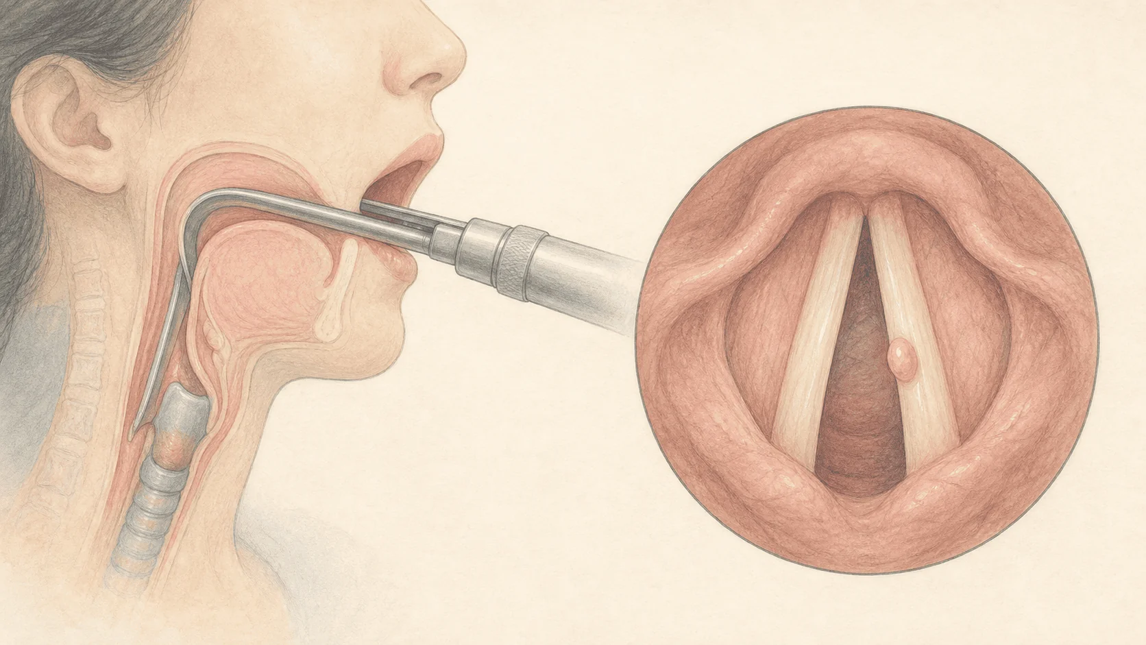
Precision laryngoscopy and voice-testing system that accurately diagnoses even subtle voice changes and minor vocal fold conditions
Thyroid Nodule Overview
Thyroid nodules are very common—about 50% of the population has them—and their prevalence increases with age. However, only about 4% of the total population actually experiences symptoms where the nodules are visible or palpable.
Lesions that develop in the thyroid gland are classified as goiters, benign thyroid tumors, and malignant thyroid tumors.
A goiter is an enlargement of the thyroid gland caused by excessive proliferation of thyroid cells, while a thyroid tumor is a mass formed when thyroid cells undergo neoplastic changes.
Thyroid Nodule Surgery System
01
Diagnosis
Check for Thyroid Abnormalities
- Thyroid Examination
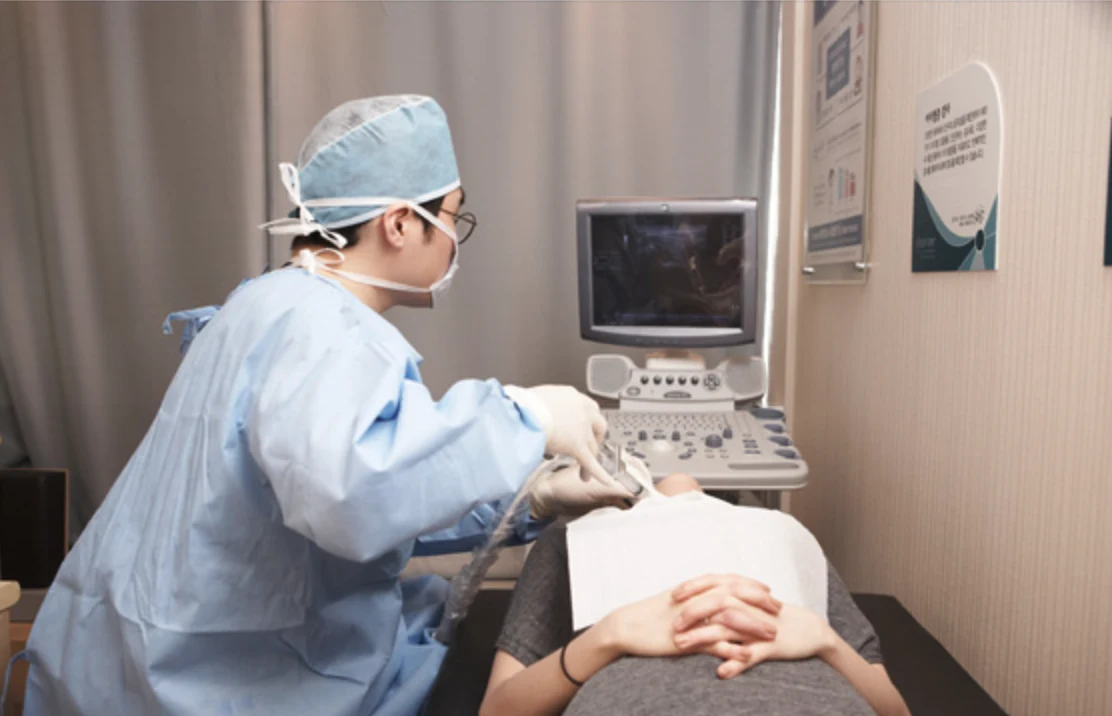
- Transabdominal Ultrasound
- Fine-Needle Aspiration Cytology
- Laryngoscopy
- Thyroid Function Tests
02
PLAN
When Surgery Is Necessary
- Computed Tomography (CT) of the Neck and Back
- Blood Test
- Urinalysis
- Electrocardiogram (ECG) Test
03
Surgery
1 DAY System
- Admission, Surgery, Discharge
- Use of Surgical Instruments for Head and Neck Tumors
04
Postoperative Care
Preventing Recurrence
- At least twice a week for the first two weeks after surgery
- Please visit the clinic depending on your condition
- Regular checkups twice a year
Thyroid Nodule Diagnosis & Testing
Of these, the fine-needle aspiration biopsy is the most important. This procedure involves inserting a thin needle into a thyroid nodule to collect a small sample of cells for examination. While it is not 100% accurate, it has virtually no side effects, is simple to perform, and is the most important test for determining whether a nodule is malignant; it is also the best method for preoperative diagnosis.
Typically, test results are available in 3–4 days, or up to a week at hospitals that take longer, but at Doori ENT Clinic, you can get your results the very next day.
- Thyroid Function Tests (Almost all results are normal)
- Ultrasound Examination
- Isotope Scintigraphy and Fine-Needle Aspiration Cytology
Neck Mass (Tumor) Surgical System
01
Diagnosis
Check for neck injuries or higher
- Uterine Examination
- Transabdominal Ultrasound
- Fine-Needle Aspiration Cytology
- Laryngoscopy
02
PLAN
When Surgery Is Necessary
- Computed Tomography (CT) of the Neck and Back
- Blood Test
- Urinalysis
- Electrocardiogram (ECG) Test
03
Surgery
1–2 DAY System
- Admission, Surgery, Discharge
- Use of Surgical Instruments for Head and Neck Tumors
04
Postoperative Care
Preventing Recurrence
- At least twice a week for the first two weeks after surgery
- Please visit the clinic depending on your condition
- Regular checkups twice a year
A lump on the neck Types
If a lump appears on your neck, you should feel the lump to check its location and firmness. An endoscopic examination of the mouth, pharynx, and larynx is performed, and the lump is evaluated using ultrasound, CT scans, and a fine-needle aspiration biopsy. A fine-needle aspiration biopsy involves inserting a thin needle into the lump to collect a small sample of cells for examination. While it is not 100% accurate, it has virtually no side effects, is a simple procedure, and is an effective method for diagnosing neck lumps prior to surgery; results are typically available within 2–3 days.
- Lymphadenitis
- Tuberculosis of the Neck
- Sebaceous Cyst
- Thyroglossal cyst
- Hemangioma
- Lymphangioma
- Metastasis of cancers of the mouth, pharynx, larynx, and other sites
- Various types of tumors, such as those in the submandibular or parotid glands, may develop